|
|
Clinical Study A – Ultrasound Data Collection Manual
|
Clinical Study A – Ultrasound Data Collection Manual
1. Introduction
|
The
following is a set of instructions for
the collection of the ultrasound images/videos during ThrombUS+ Clinical
Study A. The instructions are
addressed to the healthcare professional conventionally performing diagnostic
ultrasound scans on patients suspected of having deep vein thrombosis of the
lower limb.
|
2.
Overall data collection guidelines
|
Conventional
procedure followed on site
- Perform the conventional ultrasound diagnostic protocol for suspected deep vein thrombosis of the lower limb as you would irrespective of the ThrombUS+ Study A. - Record your findings in the conventional report as indicated by the protocol followed in your site. Additional
data collection for ThrombUS+ Study A
- For the purpose of ThrombUS+ Study A, and additionally to the conventional diagnosis report, also complete the following structured report:
*
Required - Follow the steps below to collect ultrasound images or videos as detailed. It is advisable to create a
new Ultrasound Study to record the data below for the ThrombUS+ Study.
|
|||||||||||||||||||||||||||||||||||||||||||||||||||||||||||||||||||||||||||||||||||||||||||||||
3. Ultrasound data collection steps
|
|
|||||
|
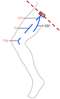
Patient
positioning and overall data collection sites
|
|
||||
|
The patient lies supine or in
Semi-Fowler's position (on their back with the head and trunk raised to
between 15 and 45 degrees), with no rotation of the pelvis. The head and shoulders
should be raised to encourage distension of the leg veins. The legs may be
tilted downwards from the head by at least 30 degrees. This helps to fill and
distend the veins, making imaging easier. |
|
||||
|
In all scanning positions: The probe should be perpendicular to
the vein. During compression ultrasound, the
probe should be compressed until the pulsatile artery compresses slightly. |
|||||
|
|
|||||
|
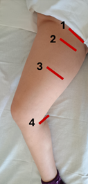
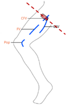
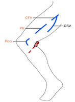
Data collection 1: Common femoral vein (CFV)
|
|
||||
|
Place the transducer in the middle of a line that connects the
pubis and the iliac spine, along the inguinal ligament, i.e. transversely to
the common femoral vein and artery. |
|||||
|
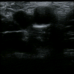
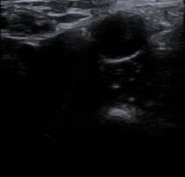
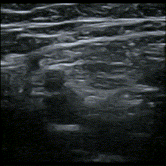
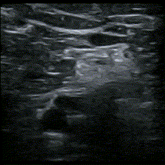
Perform a short exploratory scan around the point. Locate a frame of inadequate diagnostic quality. Save an image of inadequate
diagnostic quality. Move the transducer and locate a new frame of inadequate
diagnostic quality. Record a second image of inadequate diagnostic
quality.
|
|||||
|
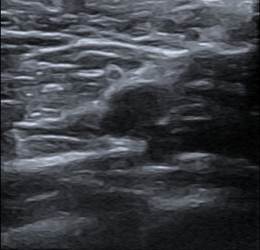
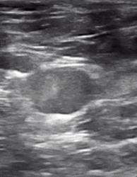
Locate the common femoral vein (CFV) and common femoral artery
(CFA). If during the conventional diagnostic exam, you have located a
site of vein incompressibility or direct thrombus visualization in this area,
choose this frame for collecting data. Start video recording. - Perform compression ultrasound Stop video recording. |
|||||
|
Total estimated duration: 5 sec |
|||||
|
Collection site 1 |
Compression video |
Image of inadequate diagnostic quality |
|||
|
|
|
|
|||
|
|
|||||
|
Data collection 2:
Great saphenous vein (GSV)
|
|||||
|
Slide the probe 1-2 cm down the patient’s leg to find where the
great saphenous vein branches off of the CFV. |
|||||
|
Perform a short exploratory scan around the point. Locate a frame of inadequate diagnostic quality. Save an image of inadequate
diagnostic quality. Move the transducer and locate a new frame of inadequate
diagnostic quality. Record a second image of inadequate diagnostic
quality.
|
|||||
|
Locate the junction of the CFV with the Great Saphenous Vein
(GSV of SV). If during the conventional diagnostic exam, you have located a
site of vein incompressibility or direct thrombus visualization in this area,
choose this frame for collecting data. Start video recording. - Perform compression ultrasound Stop video recording. |
|||||
|
Total estimated duration: 5 sec |
|||||
|
Collection site 2
|
Compression video
|
Image of inadequate diagnostic quality
|
|||
|
|
|
|
|||
|
|
|||||
|
Data collection 3:
Femoral Vein (FV)
|
|||||
|
Slide the probe a few centimeters down the patient’s leg to find
where the CFV branches into the deep femoral vein and (superficial) femoral
vein. |
|||||
|
Perform a short exploratory scan around the point. Locate a frame of inadequate diagnostic quality. Save an image of inadequate
diagnostic quality. Move the transducer and locate a new frame of inadequate
diagnostic quality. Record a second image of inadequate diagnostic
quality.
|
|||||
|
Locate the femoral vein and artery distal to the bifurcation. If during the conventional diagnostic exam, you have located a
site of vein incompressibility or direct thrombus visualization in this area,
choose this frame for collecting data. Start video recording. - Perform compression ultrasound Stop video recording. |
|||||
|
Total estimated duration: 5 sec |
|||||
|
Collection site 3
|
Compression video
|
Image of inadequate diagnostic quality
|
|||
|
|
|
|
|||
|
|
|||||
|
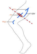
Data collection 4:
Femoral Vein (FV) – augmentation with color doppler ultrasound
|
|||||
|
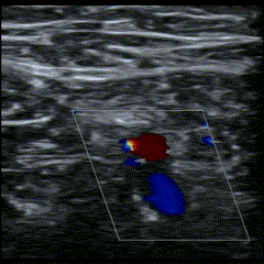
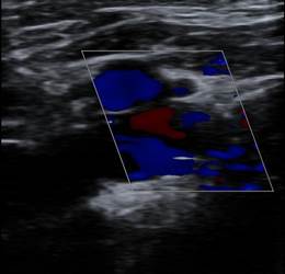
Locate the femoral vein and artery distal to the bifurcation. Switch to color doppler operation |
|||||
|
Perform a short exploratory scan around the point. Locate a frame of inadequate diagnostic quality. Save an image of inadequate
diagnostic quality. Move the transducer and locate a new frame of inadequate
diagnostic quality. Record a second image of inadequate diagnostic
quality.
|
|||||
|
Locate the femoral vein and artery distal to the bifurcation. If during the conventional diagnostic exam
you have located a site of vein incompressibility or direct thrombus
visualization in this area, choose this frame for collecting data. Start video recording. - Perform color doppler ultrasound - Squeeze the leg distal to where you are scanning (calf) Stop video recording. |
|||||
|
Total estimated duration: 5 sec |
|||||
|
Collection site 4
|
Color doppler video
|
Image of inadequate diagnostic quality
|
|||
|
|
|
|
|||
|
|
|||||
|
Data collection 5:
Popliteal Vein (PV)
|
|||||
|
Move the probe into the posterior crease of the knee and scan to
find the popliteal vein. |
|||||
|
Perform a short exploratory scan around the point. Locate a frame of inadequate diagnostic quality. Save an image of inadequate
diagnostic quality. Move the transducer and locate a new frame of inadequate
diagnostic quality. Record a second image of inadequate diagnostic
quality.
|
|||||
|
Locate the popliteal vein. If during the conventional diagnostic exam
you have located a site of vein incompressibility or direct thrombus
visualization in this area, choose this frame for collecting data. Start video recording. - Perform compression ultrasound Stop video recording. |
|||||
|
Total estimated duration: 5 sec |
|||||
|
Collection site 5
|
Compression video
|
Image of inadequate diagnostic quality
|
|||
|
|
|
|
|||
|
|
|||||
|
Data collection 6 [OPTIONAL]: |
|||||
|
If during the conventional diagnostic protocol, you happen to
visualize a clot or locate a site where vein shows pathological
incompressibility, locate this site and record image or compression video.
Total estimated duration: 3 sec |
|
||||


4. Synopsis
|
Data collection for
Study A:
1.
Short, structured report following
the table provided (page 1 or in the Annex)
2.
Point scanned: -
4 points scanned via compression ultrasound -
1 point scanned with color doppler
augmentation -
[optional] point of direct clot
visualization
3.
Ultrasound data collected -
10 images of inadequate diagnostic quality -
4 video clips of compression ultrasound (~3 min each) -
1 video clip with colour doppler augmentation (~3 min) -
[optional] image/video of direct
clot visualization 4.
Order of collected DICOM files*
-
1 - Inadequate first image of common femoral vein (CFV) -
2 - Inadequate second image of common femoral vein (CFV) -
3 - Compression video of common femoral vein (CFV) -
4 - Inadequate first image of great saphenous vein (GSV) -
5 - Inadequate second image of great saphenous vein (GSV) -
6 - Compression video of great saphenous vein (GSV) -
7 - Inadequate first image of femoral vein (FV) -
8 - Inadequate second image of femoral vein (FV) -
9 - Compression video of femoral vein (FV) -
10 - Inadequate first doppler image of femoral vein (FV) -
11 - Inadequate second doppler image of femoral vein (FV) -
12 - Color doppler video of femoral vein (FV) -
13 - Inadequate first image of popliteal vein (PV) -
14 - Inadequate second image of popliteal vein (PV) -
15 - Compression video of popliteal vein (PV) -
16 - [Optional] Image or compression video of a clot * In
case of two legs scanning, you will first acquire the DICOM files for
the left leg (L) followed by the files for the right leg (R).
|
5. Notes
|
Imaging should be conducted at the highest clinically
appropriate frequency, realizing that there is a trade-off between resolution
and beam penetration. This should usually be at a frequency of 5 MHz or
greater, with the occasional need for a lower-frequency transducer. In most
cases, a linear or curved linear transducer is preferable, but sector
scanners can be helpful for difficult patients. The potential benefits and risks of each examination
should be considered. The ALARA principle (As Low as Reasonably Achievable)
should be observed for factors that affect the acoustical output and by
considering transducer dwell time and total scanning time. Further details on
ALARA may be found in the current AIUM publication Medical Ultra-sound
Safety. Transducer preparation, cleaning, and disinfection should follow
manufacturer recommendations and be consistent with the AIUM Guidelines for
Cleaning and Preparing External- and Internal-Use Ultrasound Transducers
Between Patients, Safe Handling, and Use of Ultrasound Coupling Gel.
|
6.
References
|
1.
Jessica Ahn, Vi Dinh; et al, DVT Ultrasound
Made Easy: Step-By-Step Guide. DVT,
Ultrasound Basics, Ultrasound Tutorials POCUS 101, https://www.pocus101.com/dvt-ultrasound-made-easy-step-by-step-guide/
2.
Kakkos SK, et al. Editor's Choice - European Society for Vascular Surgery
(ESVS) 2021 Clinical Practice Guidelines on the Management of Venous
Thrombosis. Eur J Vasc Endovasc Surg. 2021 Jan;61(1):9-82. doi:
10.1016/j.ejvs.2020.09.023. 3.
Needleman L et al. Ultrasound for
Lower Extremity Deep Venous Thrombosis: Multidisciplinary Recommendations From the Society of Radiologists in Ultrasound Consensus
Conference. Circulation. 2018 Apr 3;137(14):1505-1515. doi:
10.1161/CIRCULATIONAHA.117.030687. PMID: 29610129.
4.
AIUM
Practice Parameter for the
Performance of a Peripheral Venous Ultrasound Examination. J Ultrasound Med.
2020 May;39(5):E49-E56. doi:
10.1002/jum.15263. Epub 2020 Mar 12. PMID:
32162338.
|